Dr. Prerna Agrawal, Dr. Ravi Daberao, Dr. Sarang Lambat, Dr. Prabhat Nangia, Dr. Vinay Nangia
Suraj Eye Institute, Nagpur
Case Description
A male, 10 years of age, presented on June 27th, 2022. He had a history of trauma to the left eye (LE) with a plastic ball 9 days back. He was on dorzolamide 2%, timolol 0.5 %, homatropine 2%, and prednisolone acetate 1% eye drop in LE. The best corrected visual acuity was 6/6 in the right eye (RE) and hand movement in LE. Anterior segment examination was normal in RE. Anterior segment examination of LE showed conjunctival congestion with hyphema. Intraocular pressure in RE was 14 mmHg and LE was 52 mmHg. RE Fundus examination was within normal limits. Fundus could not be visualized in LE due to hyphema. Since the IOP could not be controlled with antiglaucoma medications and was significantly elevated, a decision was made to proceed with glaucoma surgery with hyphema evacuation. He underwent a left eye trabeculectomy with MMC on 29 June 2022. Post-op day 1, bleb was flat and IOP was 23 mm Hg. Since the massage of the bleb did not work, LE bleb needling was performed on 18 July 2022. Following this, the bleb was well formed and IOP was reduced to 7 mm Hg. On fundus examination, LE showed the presence of disc edema.


Fig. 3 Colour fundus photograph of the left eye after one month also showed the presence of retinal hemorrhage (yellow arrow) in the nasal inferior quadrant.

Fig.4b Hyperreflectivity seen on the inner retina surface (white arrows).


Fig. 7 Colour fundus photograph of the left eye shows the clearing of pre-retinal hemorrhage (yellow arrow).
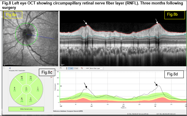

Fig. 10 Colour fundus photograph of the left eye shows further clearing of pre-retinal hemorrhage (yellow arrow).

Fig.11b There was a decrease in hyperreflectivity on the inner retina (white arrows).

Discussion
| Our patient presented to us with a history of blunt trauma with a plastic ball in his left eye. On examination, he had hyphaema in his left eye and elevated intraocular pressure (IOP) of 52 mmHg, which had not responded to medications. He underwent trabeculectomy with MMC. Following this, his IOP was still elevated at 23 mmHg. He further underwent bleb needling with the cutting of the nasal suture and movement of the 26 gauge needle under the scleral flap to break any adhesions. Postoperative the IOP was reduced to 7mmHg after bleb needling. On fundus evaluation (1 month after trabeculectomy), he had optic disc swelling in the operated eye (Fig 2 and Fig 3). The cup was not visible, and the margins were significantly blurred. The veins were tortuous. OCT scans of the RNFL showed the presence of significantly increased thickness. The macula was normal on the OCT and clinically showed exaggerated macula reflexes (Fig 2 and 3). The macula showed radiating faint yellow lines from the fovea all around, ending in yellow dots. The exact character of these changes is hard to establish (Fig. 2 and 3). He underwent neurological evaluation which was found to be normal. The macula appearance changes in character over follow-up. In September 2022, (Fig. 6) more macular reflexes are visible compared to July 2022 (Fig. 2) However, the radiating lines and the yellow dots have almost resolved completely. The character of swelling of the optic disc does not appear to be related to hypotony. Some degree of swelling of the optic disc is known to occur in association with hypotony. However, the IOP following needling was 7 mmHg. Further, there was no other sign of hypotony, especially typical hypotonic maculopathy was not seen. Optic disc swelling following blunt trauma is known to occur, though it is rare and often associated with other retinal lesions related to trauma, such as choroidal tears and decreased vision. It has been pointed out that such a swelling resolves gradually over time and often results in a degree of optic disc pallor, though there is a significant recovery of vision. In our patient, the macula showed the development of radiating folds from the fovea (Fig. 4). These persisted over follow-up but did not seem to influence the vision. There was no other sign of retinal injury. When last seen on 23rd December 2023, the visual acuity was 6/6 in BE. Over a period of several months from July to December, there was a gradual decrease in optic disc swelling. (Fig 2,3,6,7,9 and 10) and also a reduction in the RNFL swelling (Fig 5,8 and 12). While it is fortunate that the patient is recovering with masterly inactivity, it would be interesting to try to understand the cause of optic disc swelling. It is difficult to say if the optic disc fibers suffered an ischaemic episode due to blunt trauma. However, if that were the case, we might not expect a normal healthy, looking optic disc after complete recovery. It is also important to note that the vision is normal even during recovery of the optic nerve. The challenge is also in trying to understand why the resolution should take so much time. One would expect that once the pathological cause (assuming it was momentary trauma) has gone away, the resolution would be rather rapid. The delayed resolution may indicate a more significant change in the optic nerve head that eventually clinically may or may not be visible. |
ReadWise
- Mickael C. Brodsky, Kenneth J Wald, Sanford Chen et al. Protracted posttraumatic optic disc swelling. Ophthalmology. 1995; 10: 1628-1631.
- Merina Thomas, Thasarat S. Vajaranat, Ahmed A. Aref. Hyptony Maculopathy: Clinical presentation and therapeutic methods. Ophthalmol ther. 2015; 4.79-88.
Correspondence
Dr. Vinay Nangia
MS, FRCS, FRCOphth
Director
Suraj Eye Institute
Email – education@surajeye.org
QuizWise
Q.1 The following are associated with papilloedema?
A) Anterior ischemic optic neuropathy
B) Optic disc drusen
C) Hypermetropic disc
D) Intracranial mass
Q.2 Which of the following features are associated with disc edema?
A) Blurred disc margin
B) Visual field defect
C) Optic disc cupping
D) Optic atrophy
Q.3 What ophthalmic examinations will you perform on a patient suspected of or having disc edema?
A) Color vision
B) Visual acuity
C) Pupil for RAPD
D) Ocular motility
E) All of the above
Q.4 What visual field defects may present with papilledema?
A) Baring of Blind spot
B) Central scotoma
C) Enlargement of blind spot
D) Ceco-central scotoma