Dr. Sarang Lambat, Dr. Prabhat Nangia, Dr. Vinay Nangia
Suraj Eye Institute, Nagpur
Case description
A male, 70 years of age, came with a complaint of blurring of vision in both eyes for a year. He was a known hypertensive under treatment for 25 years. On examination, his BCVA was 6/12 in the RE and 6/18 in the LE. The anterior segment showed grade 2 nuclear sclerosis with grade 1 posterior subcapsular cataracts in both eyes. IOP was recorded to be 22 mmHg in both eyes. On fundus examination, RE was normal except for localised hard exudates below the fovea, and LE was normal.


He was evaluated after six months and complained of blurring in the RE. BCVA was 6/9 in the RE and 6/6 in the LE. Both eyes had clear visual axes and pseudophakia. Fundus examination revealed a further increase of hard exudates and the presence of a retinal capillary macroaneurysm.


Based on these findings, the patient was advised to undergo a focal laser to the RCM, which he got done.


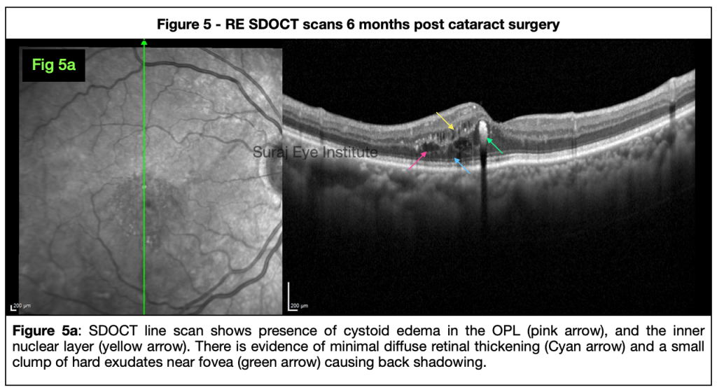
| Discussion Our case shows an increase in the retinal exudation due to the retinal capillary macro aneurysm over six months post-cataract surgery. At baseline, we could see the presence of a ring of hard exudates on fundus examination (Fig. 1). SDOCT line scan showed the presence of minimal diffuse retinal thickening with hard exudates reaching upto the fovea (Fig. 3). Only cataract surgery was planned for the patient and post-surgery he regained good VA. At six months follow-up, we could see clinically that the number of hard exudates had increased on fundus examination (Fig.4a and 4b. ) The hard exudates appear radially due to the characteristic radial arrangement pattern of the outer plexiform layer. They are seen more so involving the inferior part of the edematous area, possibly due to gravity’s effect. The aneurysms are seen to exude some amount of blood in the retinal layers, which can be seen in fig 4b. The aneurysm wall is seen to have spindle-shaped deposits (Fig 5b), which may represent the hyperreflective foci (HF) making their way through the wall of the RCM. Eventually, these HF may reach farther away from the RCM to coalesce to form the hard exudates. We planned to do a direct focal laser to the RCM. One month later, there was complete resolution of macular edema along with the involution of the RCM. (Fig. 7 and 8) The number of hard exudates and HF had also reduced significantly. Aneurysms of the vascular network are well-reported and typically involve the arterioles and the capillaries. The arteriole ones are called retinal artery macroaneurysms, and the ones that involve the capillaries are called capillary microaneurysms. Spaide et. al. described an unusual type of aneurysm, arising from capillaries in patients who did not necessarily have any other retinal vascular disease. These were typically more than 200 microns in size and were labelled as retinal capillary macroaneurysms. The capillary micro aneurysms respond well to laser photocoagulation and anti-VEGF injection. In contrast RCMs are known to show poor response to anti-VEGF injection but a good response to focal laser photocoagulation. We got similar results in our case. |
ReadWise
- Spaide RF, Barquet LA. RETINAL CAPILLARY MACROANEURYSMS. Retina. 2019 Oct;39(10):1889-1895. doi: 10.1097/IAE.0000000000002406. PMID: 30489449.
- Nadig RR, Kashyap H, Loganathan MP, Bhende M. Effect of combined laser and anti-vascular endothelial growth factor in a retinal capillary macroaneurysm. Indian J Ophthalmol. 2022 Jul;70(7):2712-2713. doi: 10.4103/ijo.IJO_581_22. PMID: 35791217; PMCID: PMC9426181.
- Rehmani AS, Banaee T, Makkouk F. Subretinal leakage of a retinal capillary macroaneurysm – a case report. BMC Ophthalmol. 2021 May 17;21(1):221. doi: 10.1186/s12886-021-01984-6. PMID: 34001046; PMCID: PMC8128355.
Correspondence
Dr. Sarang Lambat
Consultant
Vitreo-retinal services
Suraj Eye Institute
Email – education@surajeye.org
QuizWise
1. Retinal capillary microaneurysms are found in which conditions ?
A. Diabetic retinopathy
B. Retinal Vein occlusion
C. Hypertensive patients
D. All of the above
2. Typical size of a retinal capillary macroaneurysm is –
A. Less than 50 microns
B. 50 to 100 microns
C. 100 to 200 microns
D. More than 200 microns
3. Aneurysms of the retinal vasculature includes ?
A. Retinal Artery Macroaneurysm
B. Capillary Microaneurysms
C. Retinal Capillary Macroaneurysms
D. All of the above
4. Which one of the following is true about Retinal Capillary Macroaneurysms-
A. Good response to anti VEGF injections
B. Inadequate response to anti VEGF Injections
C. Poor response to laser photocoagulation
D. No response to either laser or anti VEGF injections
5. Which of the following is false related to micro aneurysms ?
A. Small micro aneurysms contain type III collagen
B. Larger ones have increased expression of MMP-9
C. Larger ones have lesser pericyte coverage
D. Larger ones have more pericyte coverage